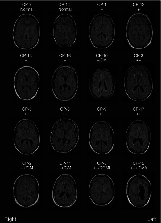
Figure 2. MRI scans .

Example axial FLAIR slices from each participant with CP imaged. Slices were selected at similar axial locations across participants except for those with cerebral malformations (CM) or deep grey matter injury (DGMI) where the slice was chosen to best display the abnormality. All participants with abnormal imaging had evidence of mild (+), moderate (++) or severe (+++) periventricular white matter injury (PVWMI) on both sides of the brain. Five participants had additional findings: CP‐2 has partial agenesis of the posterior corpus callosum (a type of cerebral malformation) and evidence of a shunt tract; CP‐10 and CP‐11 have polymicrogyria (a type of cerebral malformation); CP‐8 has injury to the deep grey matter on the left; and CP‐15 has evidence of a perinatal cerebral vascular accident (CVA).
