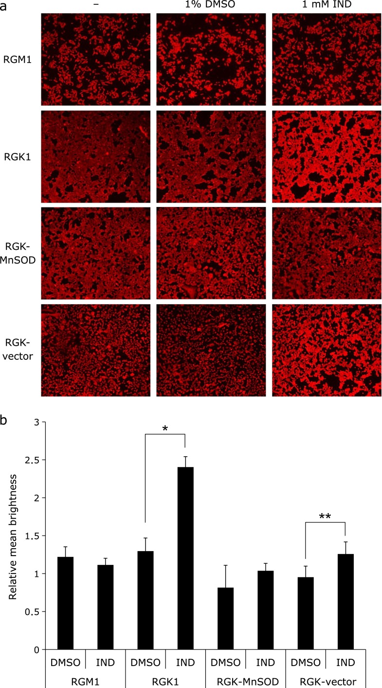
Fig. 3.
Immunohistochemical analysis of heme carrier protein 1 (HCP1) in each cell type. (a) Images of cellular immunostaining of HCP1 after exposure to 1 mM IND or 1% dimethyl sulfoxide (DMSO) or no treatment for 1 h. (b) Relative mean brightness in comparison with untreated cells was calculated in each cell type. n = 4, error bar: SD; *p<0.01, **p<0.05, Student’s t test.

