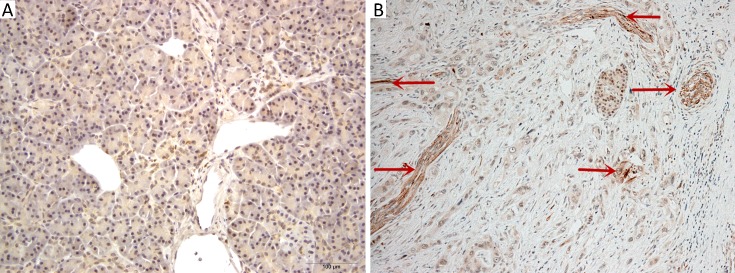
Figure 1.
Representative photomicrographs (200×) showing staining of parasympathetic nerves by expression of vesicular acetylcholine transporter (VAChT) in normal pancreatic tissue (A), and in pancreatic ductal adenocarcinoma (PDAC) (B). Scattered abnormal nerve fibers were seen in the stroma of PDAC.

