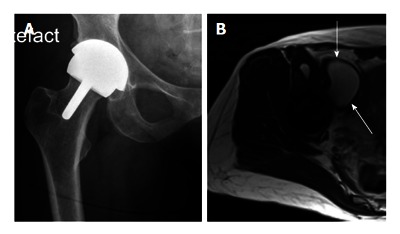
Figure 2.

Middle of the spectrum - A typical patient with moderate problems. This 58-year-old very active lady with a right hip resurfacing arthroplasty (A: X-ray AP hip) implanted 8 years ago. She has minimal symptoms and moderately raised blood metal ion levels (Cobalt 13 ppb, Chromium 7 ppb). A magnetic resonance imaging scan (B: Axial T2 weighted image) has revealed a 6 cm cystic pseudotumour anterior to the hip (arrows).
