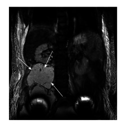
Figure 4.

A 58-year-old patient with bilateral large diameter total hip replacement metal-on-meta implanted 9-years ago, moderate hip symptoms and raised metal ion levels (Cobalt 17 ppb, Chromium 13 ppb). She presented to the general surgeons with abdominal pain and distension. Coronal magnetic resonance imaging scan (above) demonstrated a large cystic pseudotumour extending into the pelvis up to the level of the L2 vertebra and abutting the right kidney in the retroperitoneal space (arrows). The cystic pseudotumour was drained prior to surgical excision with both orthopaedic and vascular surgeons present.
