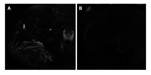
Figure 2.

Magnetic resonance image. A: Axial PD FS MR image showing increased T2 signal in the gluteus maximus, with fluid extension to the trochanteric bursa and semitendinosus bursa, fluid in the deep fascia, and subcutaneous edema. There was no hip joint effusion; B: Coronal contrast-enhanced RT FS MR image showing a central 13 cm × 5 cm area of non-enhancement in the gluteus maximus, suggestive of a hematoma. MR: Magnetic resonance.
