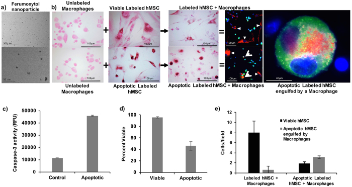
Figure 1. Macrophages phagocytose apoptotic but not viable stem cells.
(a) TEM images of ferumoxytol nanoparticles. (b) DAB-Prussian blue stains of unlabeled macrophages, viable ferumoxytol-labeled hMSCs, apoptotic ferumoxytol-labeled hMSCs, viable ferumoxytol-labeled hMSCs co-incubated with macrophages, and apoptotic ferumoxytol-labeled hMSCs co-incubated with macrophages. DAB-Prussian blue positive iron is noted as brown staining in ferumoxytol labeled hMSCs and macrophages co-incubated with the apoptotic hMSCs. Fluorescence microscopy of Rhodamine-ferumoxytol labeled hMSCs, co-incubated with anti CD68 conjugated Alexa fluor 488 antibody labeled macrophages shows red-fluorescent ferumoxytol in the cytoplasm of green macrophages in samples with apoptotic hMSC, but not viable hMSC. DAPI was used as the nucleus marker. (c) The caspase assay shows increased expression of caspase-3 in apoptotic hMSC samples compared to viable hMSC samples. Data are displayed as means and standard deviation of three samples in each group. (d) The Trypan blue exclusion test confirms a higher percentage of dead cells in apoptotic compared to viable hMSC samples. Data are displayed as means and standard error of three samples in each group. (e) Apoptotic hMSC engulfed by macrophages were counted on confocal microscopy images as the number of cells with green, red and blue color. A higher quantity of triple color positive apoptotic hMSCs engulfed by macrophages was noted in samples with apoptotic hMSCs compared to samples with viable hMSC.

