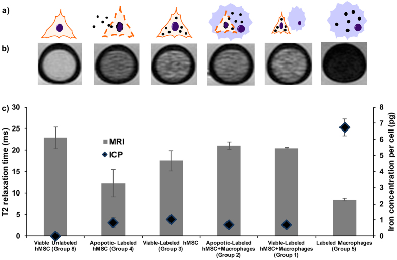
Figure 2. Iron oxide nanoparticle labeled viable MSC and iron oxide nanoparticle labeled apoptotic MSC in macrophages show no difference in MRI signal.
(a) Graphic showing different experimental groups with different distributions and compartmentalizations of iron oxide nanoparticles (black dots) in hMSCs (triangular cell symbol), and macrophages (blue round cell symbol). (b) Axial T2 weighted (TE/TR = 30 ms/3000 ms) images of viable unlabeled hMSCs, viable/apoptotic labeled hMSCs, viable/apoptotic labeled hMSCs co-incubated with macrophages, and labeled macrophages in Ficoll in NMR test tubes. (c) Corresponding T2-relaxation times (grey bars) and iron content, quantified by ICP-OES (blue diamonds). Data are displayed as means and standard error of triplicate samples in each group.

