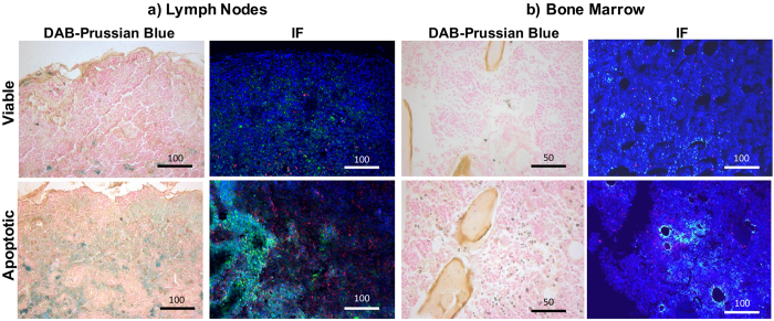
Figure 4. Migration of iron-loaded macrophages into surrounding bone marrow and regional lymph nodes.
DAB-Prussian blue and CD68 immunofluorescence stains of (a) lymph nodes and (b) bone marrow of mice after transplantation of viable or apoptotic hMSC transplants. The higher amount of brown colored cells in the DAB-Prussian blue stains in apoptotic transplants suggests macrophages migration into both bone marrow and lymph nodes. Accordingly, immunofluorescence stains show a higher amount of ferumoxytol (green) and macrophages (red) in the bone marrow and popliteal lymph nodes of knee joints with apoptotic transplants.

