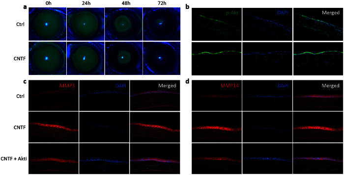
Figure 5. Akt contributes to the elevation of MMP3 and MMP14 in vivo.
Normal mice were injected subconjunctivally with 50 ng CNTF or PBS (control group) after the scrape of corneal epithelium. Six mice were used in each group (n = 6). Immunofluorescence intensity of p-Akt, MMP3 and MMP14 was compared between groups in the regenerated region of corneal epithelium at 48 hour post scratching. Representative figures are shown. (a) The defect area of corneal epithelium was evaluated after 24, 48 and 72 hours by 0.25% fluorescein sodium under a BQ900 slit lamp. ‘h’ indicates hours. (b) Immunofluorescence staining was carried out to compare the expression of p-Akt. The right column is the merged picture of left column and middle column. (c,d) For Akt inhibition, the mice were injected with the Akt inhibitor 24 hours before CNTF injection. Immunofluorescence staining was carried out to compare the expression of MMP3 (c) and MMP14 (d) between groups.

