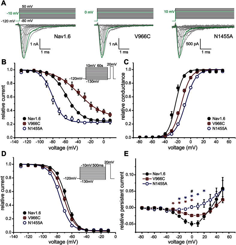
Figure 4. SI of Nav 1.6 is modified by the mutations Nav1.6/V966C and Nav1.6/N1455A.
(A) Representative current traces of Nav1.6 (left panel), the mutation Nav1.6/V966C (middle panel) and the mutation Nav1.6/N1455A (right panel) expressed in ND7-cells. For each channel the maximal inward current and corresponding potential is colored in green. (B) Steady-state SI is enhanced by the Nav1.6/N1455A mutation (open blue circles, V1/2 = −81.9 ± 2.5 mV, n = 4) and decreased by the Nav1.6/V966C mutation (crossed red circles, V1/2 = −33.8 ± 5.1 mV, n = 5) compared to Nav1.6 WT (filled black circles, V1/2 = −59.6 ± 1.5 mV, n = 13). (C) Normalized conductance–voltage relationship is shifted to more depolarized potentials for both Nav1.6 mutants whereas (D) the V1/2 of steady-state fast inactivation is slightly hyperpolarized. (E) Nav1.6 WT (filled black circles) and the mutant Nav1.6/V996C (crossed red circles) display persistent currents but the mutant Nav1.6/N1455A (open blue circles) does not. Data shown in E were recorded in the presence of β4-peptide and 50 nM ATX-II and were significantly different for Nav1.6 WT and Nav1.6/N1455A (blue*), for Nav1.6 WT and Nav1.6/V966C (red*) and for Nav1.6/N1455A and Nav1.6/V966C (black#), p < 0.05.

