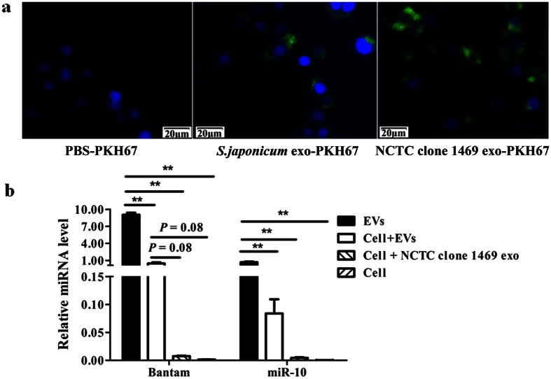
Figure 6. Uptake of S. japonicum EVs by murine liver cells.
(a) Uptake of S. japonicum EVs by normal mouse liver cells detected using fluorescence microscopy. Approximately 10 μg of PKH67-labelled S. japonicum EVs, NCTC clone 1469 exosomes, and a PKH67-PBS control were added to the cells. Uptake of the labeled exosomes by NCTC clone 1469 cells was detected using fluorescence microscopy. Nuclei were stained with DAPI (blue). (b) qRT-PCR analysis of S. japonicum EV associated miRNAs in treated cells. Results represent mean ± SEM from triplicate experiments. *P ≤ 0.05 and **P ≤ 0.01 (Tukey’s test, one-way ANOVA).

