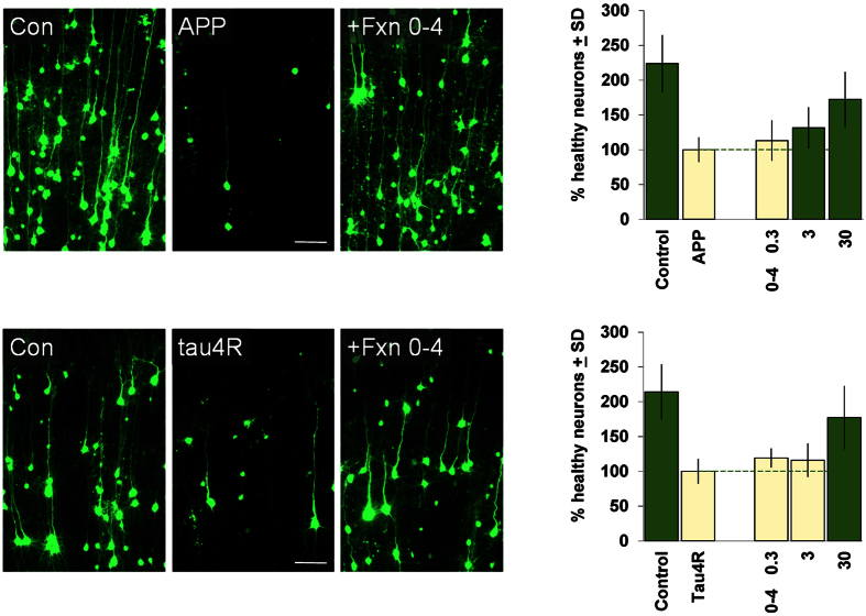
Figure 2. Neuroprotective activity of Fraction 0–4 in brain slice assays for neurodegeneration induced by APP and tau.
Left, example photomicrographs showing cortical neurons transfected with YFP only (“Con”) or with YFP plus a human WT amyloid precursor protein (“APP”) or a human tau4R0N (“tau4R”) expression construct as indicated. Overt neurodegeneration and cell loss driven by either APP or tau4R by 3 days after transfection (compare middle to left panels) could be rescued by treatment with 30 μM Fraction 0–4 (right panels). Right, Concentration-response relations for Fraction 0–4 in the brain slice APP and tau4R assays as indicated. Averages of 3 and 4 independent runs are shown for APP and tau4R, respectively, with the negative-control conditions (treated with DMSO only) set to 100%. For both graphs, dark green bars denote statistically significant differences with respect to the respective APP or tau4R negative-controls by ANOVA followed by Dunnett’s post hoc comparison test at the 0.05 confidence level.

