FIGURE 1.
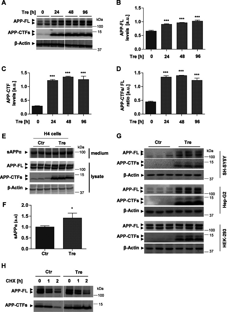
Trehalose induces accumulation of APP and APP-CTFs. A, Western immunoblot of APP and APP-CTFs. H4 cells were incubated in the presence or absence of 100 mm trehalose (Tre) for the indicated time periods. Cell lysates were subjected to SDS-PAGE and Western immunoblotting. β-Actin was used as a loading control. B–D, quantification of APP-FL (B), APP-CTFs (C), and the ratio APP-CTFs/FL (D) by ECL imaging. Values represent means ± S.D. of six biological replicates. E, H4 cells were incubated in the presence or absence of 100 mm trehalose (Tre) for 24 h. Soluble APPα in conditioned media and APP-FL and APP-CTFs in cell lysates were analyzed by Western immunoblotting. F, quantification of soluble APPα in media by ECL imaging. Values represent means ± S.D. of three biological replicates. G, the indicated cell types (SH-SY5Y, Hep-G2, and HEK-293) were incubated in the presence or absence of 100 mm trehalose for 24 h. Cells were lysed, and proteins were detected by Western immunoblotting. H, H4 cells were incubated in the presence or absence of 100 mm trehalose (Tre) for 2 h. After addition of cycloheximide (CHX; 20 μg/ml), cells were further incubated for the indicated time periods. Cellular membranes were isolated, and APP-FL and APP-CTFs detected by Western immunoblotting. Error bars represent S.D. *, p < 0.05; ***, p < 0.001. a.u., arbitrary units; sAPP, soluble APP; Ctr, control.

