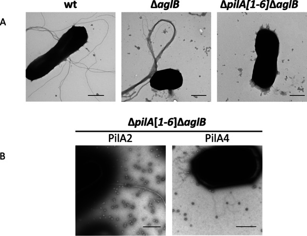
FIGURE 1.

Glycosylation differentially affects pilus structures and interactions. A, TEM of whole cells of wild-type (wt), ΔaglB, and ΔpilA[1–6]ΔaglB strains transformed with pTA963; B, pTA963 expressing PilA2 or PilA4. WT cells, which have both pili and flagella on the surface (as determined by MS) (Fig. 2) (37), form long single filaments, although the pili on ΔaglB cells (as determined by MS (Fig. 2)) form long thick bundles. The ΔpilA[1–6]ΔaglB strain lacks pili and flagella. Although PilA2 forms long bundles of pili when expressed in the ΔpilA[1–6]ΔaglB strain, PilA4 has individual pili that unlike WT are curly when expressed in this background. Bars, 500 nm (n ≥2).
