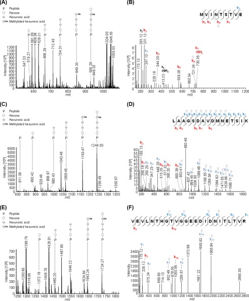
FIGURE 2.
Mass spectrometric identification of novel N-glycopeptides from FlgA1 and FlgA2. Peptides of GluC or trypsin-digested proteins from CsCl density gradients obtained from WT cells were analyzed by MS applying IS-CID (80 V). The N-glycan composition could be analyzed by a series of neutral losses in the MS1 spectra (A, C, and E) and resulted in the identification of a pentasaccharide composed of hexose (○), hexuronic acid (♢), and a methylated hexuronic acid (♢Me). The corresponding N-glycopeptide sequence was analyzed by further fragmentation (HCD) of a precursor ion corresponding to the peptide (P) + hexose and led to the identification of MVIN@TSTVE from FlgA1 (B, precursor m/z 578.27, charge 2) as well as LAAGSDAVDMN@ETSIK (D, precursor m/z 892.42, charge 2) and VEVLNTHGTVGGEEDIDN@ITLTVR (F, precursor m/z 1372.19, charge 2). @ corresponds to the N-glycosite. The observed b-ions (red) and y-ions (blue) are indicated in the spectrum as well as the fragmentation scheme, and the circle indicates the presence of a hexose attached to the peptide or fragment ion. Some peaks (slanted parallel bars) are not shown with their full intensity for better visualization (n = 1).

