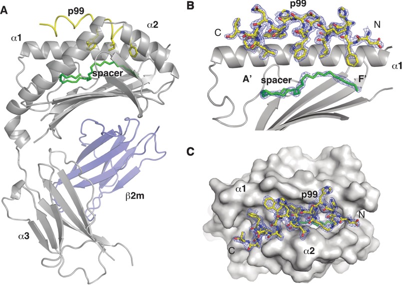
FIGURE 1.
Crystal structure of the CD1d-p99 complex. A, overall view of the mCD1d-p99 complex with the CD1d chain in gray, β2m in blue, the p99 peptide in yellow, and the spacer in green. The p99 peptide binds in the antigen-binding groove, between the α1 and α2 helices and above the spacer filling the remaining of the cavity. B, side view of the antigen-binding groove with the α2 helix removed for clarity. 2 Fo − Fc density at 1.0 σ is shown for the peptide and spacer molecule. C, top view of the CD1d-p99 complex with the CD1d molecule shown as a surface and the 2Fo − Fc density at 1.0σ for the peptide.

