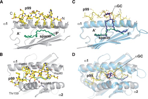
FIGURE 2.
Binding mode of the p99 peptide to CD1d. A, side view of the antigen-binding groove with the α2 helix removed for clarity. The residues composing the CD1d-binding motif are labeled, highlighting how they are all oriented toward the cavity of the A′ and F′ pockets. B, top view of the antigen-binding groove with highlighted the polar contacts between CD1d and the p99 peptide. C, superposition of the CD1d-p99 and CD1d-α-GalCer structure. In the side view, α2 helix was removed for clarity. Note how the peptide and α-GalCer overlap and protrude to similar extents from the antigen-binding groove. D, top view of the antigen-binding groove with the superposition of the CD1d-p99 and CD1d-α-GalCer structures.

