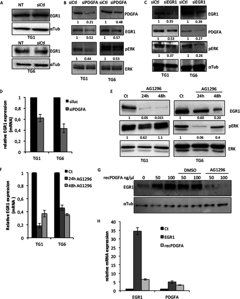
FIGURE 7.
PDGFA signaling contributes to the maintenance of EGR1 expression in GSC. A, immunoblotting showing the unaffected EGR1 expression in TG1 and TG6 cells transfected with a non-relevant control siRNA (siCtl). This expression is compared with the basal EGR1 expression in untreated cells (NT). A specific antibody to α-tubulin was used as loading control. B and C, immunoblotting showing the effects of siRNA-mediated PDGFA- and EGR1-specific inhibition (siPDGFA and siEGR1) on phosphorylation of ERK in TG1 and TG6 cells. Antibodies specific to total ERK or α-tubulin were used as loading control. D, QPCR showing EGR1 mRNA levels in TG1 and TG6 in response to siPDGFA or siLuc treatment. E and F, immunoblotting (E) and qPCR (F) showing the effect of PDGFR chemical inhibitor AG1296 on EGR1 expression and ERK phosphorylation in TG1 and TG6 cells. Cells were treated for 24 or 48 h. A specific antibody to total ERK was used as loading control. Quantification is shown below each blot. G, immunoblotting showing EGR1 expression in untreated cells or cells treated with increasing amounts of recPDGFA in the presence of DMSO, used as control, or AG1296. A specific antibody to α-tubulin was used as a loading control. H, relative expression of EGR1 and PDGFA in untreated (Ct) or EGR1-expressing (EGR1) cells or cells treated with recPDGFA (recPDGFA). Error bars, S.E.

