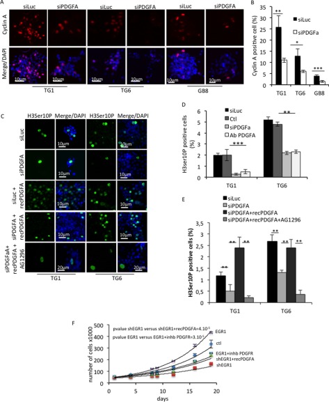
FIGURE 8.
PDGFA is involved in the GSC proliferation. A, immunofluorescence staining showing cyclin A expression in TG1, TG6, and GB8 cells transfected with a control siRNA (siLuc) or a siRNA to PDGFA (siPDGFA). Nuclei were stained with DAPI. Scale bars are shown. B, quantification of cyclin A-positive cells in each primary culture. Five independent fields were counted. *, p < 0.05; **, p < 0.01; ***, p < 0.001; Student's t test). C, immunofluorescence staining showing mitotic cells assessed by the Ser-10 phosphohistone 3 expression in TG1 and TG6 cells cultured in defined medium. Cells were transiently transfected with a control siRNA (siLuc) or a PDGFA-targeting siRNA (siPDGFA) in the presence or absence of recombinant PDGFA protein (recPDGFa) to rescue the PDGFA signaling. When indicated, cells were treated with AG1296. Nuclei were stained with DAPI. Scale bars are shown. D, quantification of mitotic cells by counting the Ser-10 phosphohistone 3-positive cells for untreated (Ctl) TG1 and TG6 cells or transiently transfected with a non-relevant siRNA (siCtl), siPDGFA, or treated with a PDGFA-blocking antibody (Ab PDGFA) (**, p < 0.01; Student's t test). E, quantification of mitotic cells assessed by Ser-10 phosphohistone 3 staining in TG1 and TG6 treated as shown in C by counting the Ser-10 phosphohistone 3-positive cells for TG1 and TG6 cells with siLuc or siPDGFA or treated with a blocking PDGFA antibody (**, p < 0.01; Student's t test). F, cell proliferation of TG6 cells efficiently transduced with either an EGR1 expression vector or a specific shRNA and compared with the control condition. When indicated, cells were treated with a PDGFR chemical inhibitor or a recombinant PDGFA protein. The count was performed over 20 days. Error bars, S.E.

