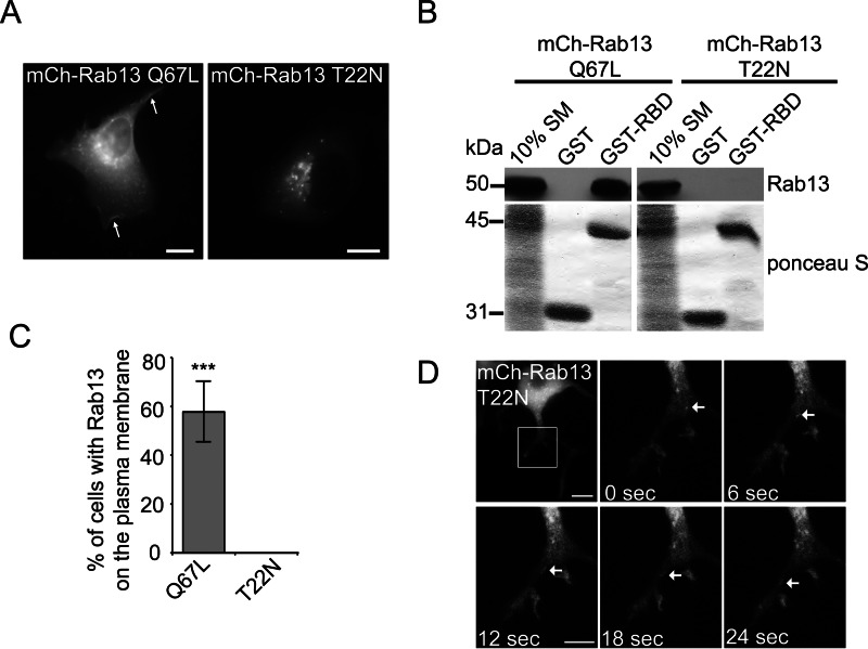
FIGURE 1.
Inactive Rab13 traffics on vesicles. A, MCF10A cells expressing mCh-Rab13 Q67L or mCh-Rab13 T22N were imaged live. The arrows illustrate localization on the plasma membrane. Scale bars = 10 μm. B, the GST-Rab-binding domain (RBD) or GST alone, bound to glutathione-Sepharose, was incubated with HEK-293T cell lysates expressing mCh-Rab13 Q67L or T22N. Ponceau S staining revealed the level of GST-Rab-binding domain, whereas specifically bound Rab13 was detected by blot. Starting material (SM) equals 10% of the lysate used per condition. C, the percentage of transfected cells with mCh-Rab13 constructs on the plasma membrane. Data are mean ± S.D., measuring a minimum of 20 cells/experiment from a minimum of three independent experiments (Student's t test; ***, p < 0.001). D, PC12 cells differentiated for 24 h by addition of 50 ng/ml NGF and expressing mCh-Rab13 T22N were imaged live. The boxed region is magnified. The arrows follow a vesicle trafficking along a neurite. Scale bar = 10 μm (top panel) and 5 μm (magnified panel).

