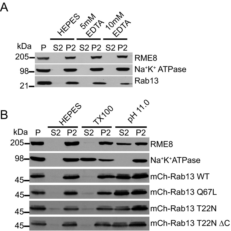
FIGURE 7.
Rab13 associates in a protein complex independent of nucleotide status. A, HEK-293T cell homogenates in HEPES buffer were spun for 30 min at 200,000 × g, and equal protein aliquots of the resulting pellet (P) were resuspended in ice-cold HEPES buffer with or without EDTA at the indicated concentrations. After 15 min of incubation, the samples were spun for 30 min at 200,000 × g, and the resulting supernatant (S2) and pellets (P2) were analyzed by Western blotting using the indicated antibodies. B, HEK-293T cells were left untransfected or were transfected with various mCh-Rab13 constructs as indicated. Cell homogenates in HEPES buffer were spun for 30 min at 200,000 × g, and equal protein aliquots of the resulting pellet were resuspended in ice-cold HEPES buffer with or without 1% Triton X-100 (TX100) or NaCO3 at pH 11.0. After 15 min of incubation, the samples were spun for 30 min at 200,000 × g, and the resulting supernatant (S2) and pellets (P2) were analyzed by Western blotting using the indicated antibodies for the first and second panels and an antibody against Rab13 to detect the various Rab13 constructs.

