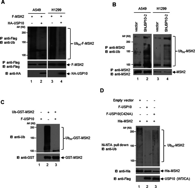
FIGURE 3.
USP10 deubiquitinates MSH2. A, USP10 deubiquitinates MSH2 in vivo. A549 and H1299 cells were transfected with the indicated plasmids. Twelve hours prior to harvest, 10 μm MG132 was added to the cells. F-MSH2 was IP-ed by anti-Flag antibodies. The immunoprecipitates were then subjected to the anti-Ub Western blotting analysis (upper panels). The blot was stripped and reprobed with anti-Flag antibodies (middle panels). The expression of HA-USP10 was detected by the anti-HA Western blotting analysis (lower panels). B, knockdown of USP10 increases MSH2 ubiquitination in vivo. USP10 was stably transfected by vector control or shRNA against USP10 (shUSP10–2) in A549 and H1299. The cells were treated overnight with 25 μm MG132 prior to harvest. MSH2 was IP-ed by anti-MSH2 antibodies. The immunoprecipitates were then subjected to an anti-Ub Western blotting analysis (upper panels). Ten percent of the above IP-ed samples was subjected to the anti-MSH2 Western blotting analysis for determining IP efficiencies (lower panels). C, USP10 deubiquitinates ubiquitinated MSH2 in vitro. The Ub-GST-MSH2 protein was prepared as described in the “Experimental Procedures.” Ub-GST-MSH2 was incubated in the absence or presence of F-USP10 protein purified from 293T cells in the deubiquitination buffer (50 mm Tris-HCl, pH 8.0, 50 mm NaCl, 1 mm EDTA, 10 mm DTT, 5% glycerol) at 37 °C for 1 h. The reaction was resolved on SDS-PAGE followed by an anti-Ub Western analysis (upper panel). The amount of GST-MSH2 was examined by an anti-GST Western blotting analysis (lower panel). D, USP10-WT, but not USP10-(C424A) mutant, deubiquitinates MSH2 in vivo. 293T cells were transfected with indicated constructs. His-MSH2 was pulled down with Ni-NTA-agarose beads under denaturing conditions followed by anti-Ub Western blotting analysis (upper panel). The expression of His-MSH2 and USP10-WT and the USP10-CA mutant was examined by anti-His (middle panel) and anti-Flag (bottom panel) Western blotting analyses.

