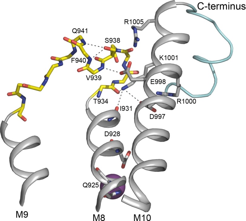
FIGURE 7.
Structural relations of L8-9 containing the potential PKA site Ser-938. Shown is the relevant part of the structure of the Na+-bound E1 form of Na+,K+-ATPase (Protein Data Bank code 3WGV) (8) viewed from the side along the membrane surface. Depicted is the extensive bonding network that connects L8-9 (yellow) harboring the potential PKA site at Ser-938 with M10 (gray), the C terminus (light blue), and the Na+ binding segment M8 (gray). The residues studied by mutagenesis are depicted as sticks (Ser-938, Asp-997, Glu-998, Arg-1000, Lys-1001, and Arg-1005). Stick representation of backbone atoms (blue nitrogen and red oxygen atoms) is shown for the entire L8-9. In addition, Gln-925 and Asp-928 known to contribute to the binding of the Na+ ion at site III are shown as sticks as are Ile-931 and Thr-934 that interact with Lys-1001 in M10. The Na+ ion bound at site III is depicted as a large purple sphere. The figure was prepared using PyMOL.

