
Fig. 3.
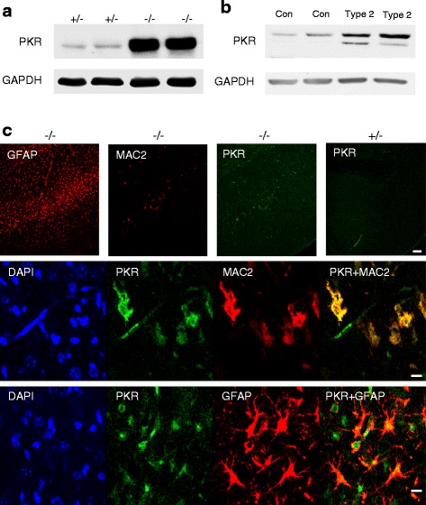
Elevation of PKR (Eif2ak2) in Gba −/− mice. a Western blot of homogenates (150 μg of protein) from the cortex of 21-day-old Gba −/− mice (n = 5). GAPDH was used as loading control. b Western blot of homogenates (50 μg of protein) from the cerebellum of two human patients who succumbed to type 2 GD compared to an age-matched control brain. c PKR levels in microglia/macrophages and in astrocytes in the Gba −/− brain. Immunofluorescence of cortical layer V in 16-day-old Gba −/− using anti-PKR (green), anti-MAC2 (red), or anti-GFAP (red) antibodies. Upper panel PKR staining is located in pathological areas as shown by staining of both MAC2 and GFAP. Scale bar, 100 μm. Middle and lower panels Double immunofluorescence of 16-day-old Gba −/− mice using either anti-PKR and anti-MAC2 (middle panel) or anti-PKR and anti-GFAP (lower panel) antibodies. PKR is in green, MAC2 and GFAP are in red, and areas of overlap are indicated on the right. Scale bar, 10 μm. Results are representative of three experiments