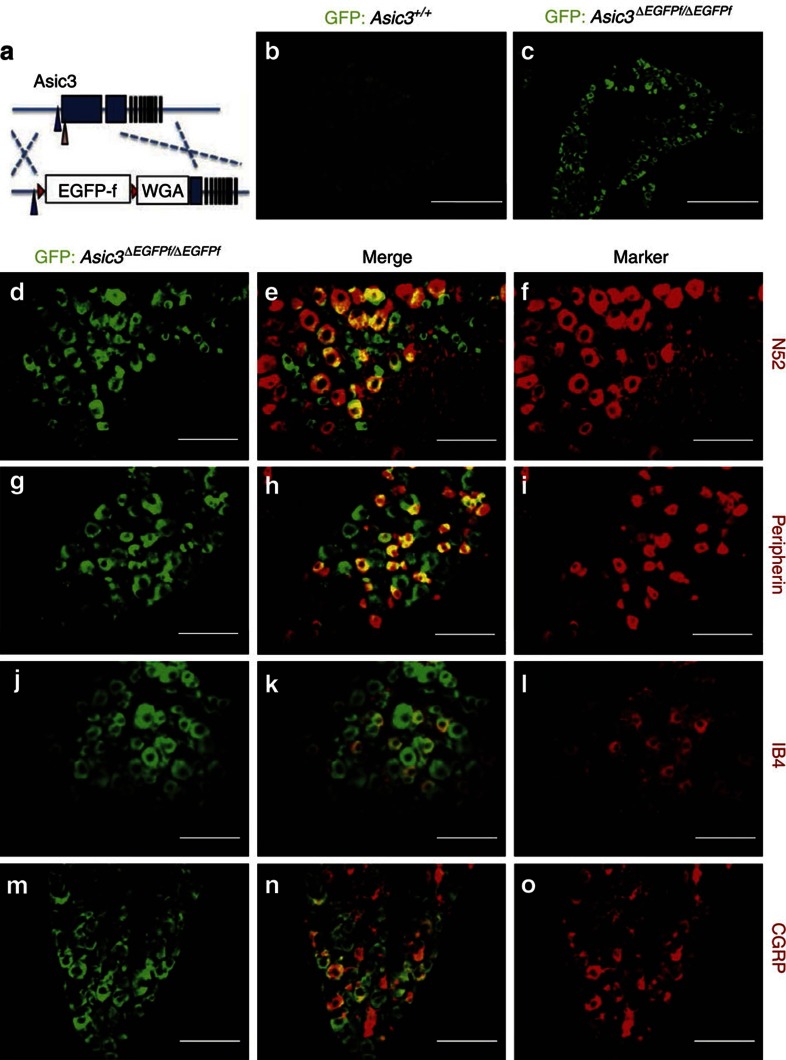
Figure 1. Immunofluorescence staining of GFP in ASIC3-expressing DRG neurons in Asic3-KO/eGFP-f-KI (Asic3ΔEGFPf/EGFPf) mice.
(a) Illustration of the EGFPffloxed-WGA construct targeted in Accn3 allele in mice. Blue and pink arrowheads indicate the position of the transcription and translation start sites of mouse Accn3, respectively. Red triangles represent the loxP sites. Blue boxes indicate the exons. (b) GFP signal was very low in wild-type lumbar DRG (scale bar, 50 μm). (c) Expression of GFP was much higher in the Asic3ΔEGFPf/EGFPf lumbar DRG (scale bar, 50 μm). (d–o) Co-localization of GFP signals with: (d–f) myelinated neuron marker N52, (g–i) small nociceptor marker peripherin, (j–l) non-peptidergic nociceptor marker IB4 and (m–o) peptidergic nociceptor marker CGRP in the Asic3ΔEGFPf/EGFPf lumbar DRG are illustrated (scale bar, 100 μm).

