
Fig. 6.
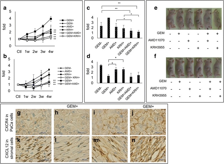
In vivo tumorigenicity of GEM-R PaCa cells and inhibition by CXCR4 antagonists. The growth of subcutaneous implanted GEM-S and GEM-R MIA PaCa-2 cells in nude mice. Mice were divided into 6 groups for each treatment: group I was not given any drugs, group II was given GEM, group III was given AMD11070, group IV was given KRH3955, groupV was given GEM and AMD11070 and groupVI was given GEM and KRH3955. The measurements of tumor volumes after implantation of a GEM-R or b GEM-S in each treatment group were plotted 4 weeks after beginning of the treatment. Values are expressed as means ± SD. Multiple comparisons were performed by using one-way ANOVA followed by Dunnett’s test, **, P < 0.01; *, P < 0.05 versus control (group II in GEM-R group, group I in GEM-S group at 4 weeks). The differences of tumor volumes after implantation of GEM-R (c) or GEM-S (d) were measured and photos showed representative results of GEM-R (e) and GEM-S (f) in each treatment group 4 weeks after beginning of the treatment. Values are expressed as means ± SD. Multiple comparisons were performed by using one-way ANOVA followed by the Bonferroni test, **, P < 0.01, *, P < 0.05 among all groups. Expression of CXCR4 protein determined by immunohistochemical staining (brown, CXCR4 protein, blue, nucleus) in implanted PaCa tumor: g GEM-S, h GEM-S treated with GEM, (i) GEM-R, (j) GEM-R treated with GEM. The secretion of CXCL12 from stromal cells around PaCa cells by immunohistochemical staining (brown, CXCL12 protein, blue, nucleus) in stromal cells, k stromal cells around GEM-S, l around GEM-S treated with GEM, m around GEM-R, n around GEM-R treated with GEM. Original magnification, ×1000, scale bars, 20 μm