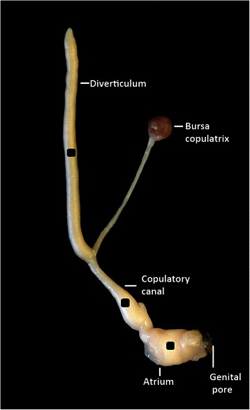
Fig. 1.

The part of the reproductive system of C. aspersum that was used in this study. This includes the genital pore where the partner’s spermatophore enters; atrium; copulatory canal; the bursa copulatrix which is responsible for sperm digestion; the diverticulum which receives the spermatophore. This is the preparation used for each experimental trial. The three black squares (±2 mm2) of electrical tape glued onto diverticulum, copulatory canal and atrium were used as markers. The position of these markers was recorded with a webcam, then tracked with DLTdv5 marker tracking software to measure the response of the preparation to each species mucous extracts added to the saline bath (see Additional file 1: Movie 1). Not illustrated: the small dish containing 2 ml saline solution where the preparation was placed and the pins on both sides along the length of the diverticulum, in the Sylgard base of the small dish, to make the measurements comparable
