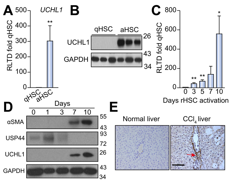
Fig. 2. UCHL1 expression is significantly elevated in activated (aHSC) vs. quiescent (qHSC) rat stellate cells.
(A) UCHL1 mRNA expression levels were quantified by qRT-PCR and expressed as relative level of transcriptional difference (RLTD) of activated rat HSC (day 10) compared with quiescent (day 0) n = minimum of 3 animals per group. (B) Western blots for UCHL1 expression in quiescent (n = 3) and day 10 activated rat HSC (n = 3). (C) UCHL1 mRNA expression in rat HSC (rHSC) harvested at days 0–10 of culture and results are expressed as RLTD (day 0 HSC). (D) Western blot for αSMA, USP44, UCHL1, and GAPDH expression in rat HSC harvested at days 0–10 of culture. Molecular weight markers shown on right panel. (E) Representative photomicrographs of UCHL1 stained liver sections from rats treated with olive oil vehicle (left panel) or CCl4 injured for 4 weeks (right panel), n = 3 independent experiments. Statistical analysis between quiescent and activated HSC were compared using two-tailed unpaired students t test *p <0.05; **p <0.01.

