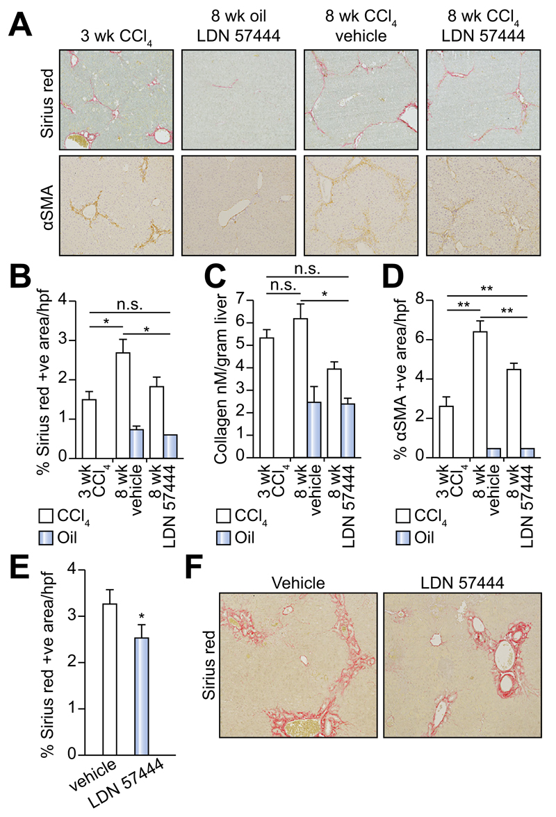
Fig. 5. Inhibition of UCHL1 reduces fibrogenesis in two liver fibrosis models.
(A) Representative photomicrographs of Sirius red and αSMA stained liver sections from 3 week CCl4, 8 week oil/LDN 57444, 8 week CCl4/vehicle and CCl4/LDN 57444 treated mice. (B) Fibrosis densitometry of Sirius red stained liver sections. (C) Liver collagen content as assessed by hydroxyproline assay. (D) Average mean% area of αSMA stained liver sections (n = 5 per group). (E) Fibrosis densitometry and (F) representative photomicrographs of Sirius red stained liver sections from BDL control or LDN 57444 treated mice (n = 8 control and 7 LDN 57444). Multicomparison analysis was performed using two-way repeated measures ANOVA between all groups p <0.05 considered significant; followed by Tukeys post-tests between individual groups where *p <0.05; **p <0.01.

