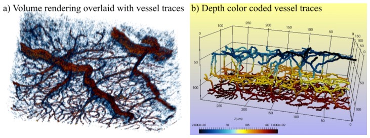
Fig. 5.
Results of user-guided retinal vessel tracing obtained with the Simple Neurite Tracker plugin of ImageJ software and showcased with ParaVIEW software. a) Volume rendering of the vessels overlaid with the vessel traces. b) 3D skeleton visualization of the traced vessels. Three vascular layers are visible: retinal vessels (blue), inner capillary plexus (yellow) and outer capillary plexus (red), as well as vessels connecting these layers. We have provided more detailed presentation of b) in Visualization 1 (77.6MB, AVI) .

