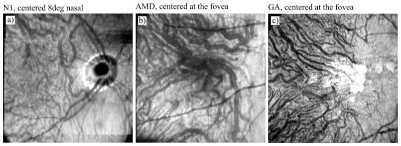
Fig. 8.
Overview of the choroidal vascular layers - en face projection of Haller's layer obtained from the intensity data acquired in: a) a normal eye (subject N1), b) an age related-macular degeneration case, and c) a geographic atrophy (GA) case. Image locations: 8° nasal from the fovea in N1, and at the fovea in the AMD and GA cases. Image sizes: 9 x 9 mm.

