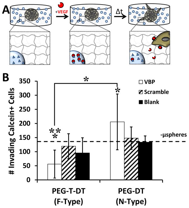
Figure 5.
Influence of VBP microspheres on iPSC-EC sprouting behavior in hydrogels. A:Schematic demonstrating iPSC-EC sprouting away from cell-dense sphere into surrounding synthetic hydrogel with encapsulated VBP microspheres. B: iPSC-EC sprouting quantified as the number of invading Calcein+ cells for each condition. Condition with no microspheres (-μspheres) in the presence of VEGF is shown with a dashed line. Two-way analysis of variance was performed (microsphere peptide identity p-value> 0.05, microsphere crosslink type p-value= 0.002, interaction p-value= 0.016) with post-hoc Student’s t-test. Statistical significance for Student’s t-test denoted compared to Scramble (**) and no microsphere (*) conditions or between conditions in brackets at α = 0.05). Data is presented as mean + /− standard deviation for eight replicates per condition

