Figure 3.
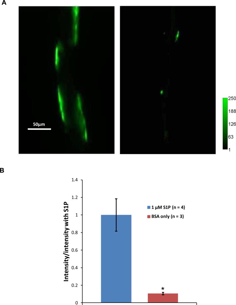
(A) Images of fluorescently labeled heparan sulfate in a post-capillary venule pretreated with 1 μM S1P (left) and a vessel without S1P (right). (B) Comparison of the intensity of the fluorescently labeled heparan sulfate in 4 vessels with S1P and that in 3 vessels without S1P. * p < 0.001.

