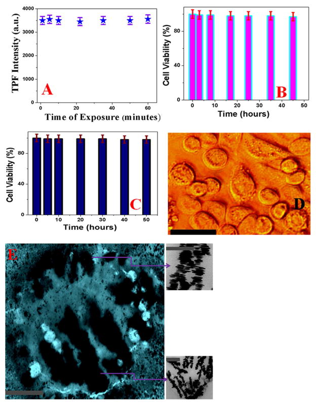
Figure 5.
A) Plot shows the photo-stability of TPF intensity at 700 nm from anti-GPC3 antibody attached DNA-mediated gold nanoprism assembly even after an hour of exposure with 1100 nm light. B) Bar plot shows very good biocompatibility even after 48 hours incubation of anti-GPC3 antibody attached DNA-mediated triangular gold nanoprisms assembly against human Hep G2 liver cancer cells. After two days of incubation with human Hep G2 liver cancer cells, we have observed about 98% cell viability. C) Bar plot shows very good biocompatibility even after 50 hours incubation of anti-GPC3 antibody attached DNA-mediated triangular gold nanoprisms assembly against HaCaT normal skin cells. After more than two days of incubation with human HaCaT normal skin cells, we have observed about 97% cell viability. D) Bright-field inverted microscopic images of human Hep G2 liver cancer cells after 48 hours of incubation with anti-GPC3 antibody attached DNA-mediated triangular gold nanoprisms assembly. Reported data indicate that cancer cells are alive even after two days of incubation. (scale bar = 20 μm) E) TEM image shows single human Hep G2 liver cancer cell is conjugated with anti-GPC3 antibody attached nanoprisms assembly (scale bar = 1 μm). Inserted picture shows the morphology of anti-GPC3 antibody attached nanoprisms assembly on Hep G2 liver cancer cell surface (scale bar = 200 nm).

