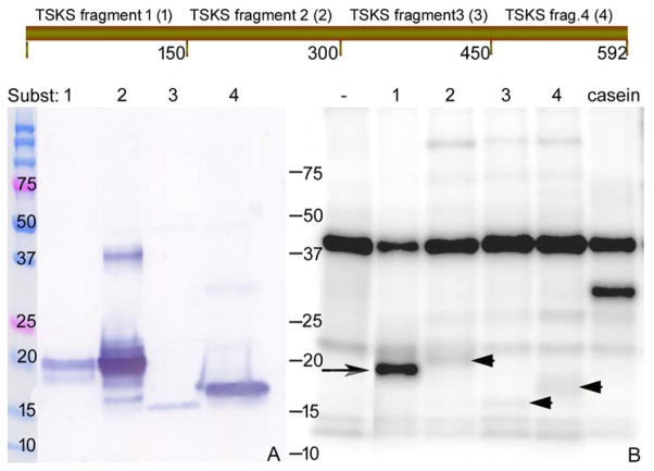
Figure 7.
Identification of a TSKS domain involved in phosphorylation by hTSSK2. hTSKS isoform1 was divided into 4 fragments of ~150 amino acids as shown and recombinant fragments 1–4 were generated with a His-tag at each C-terminus. (A) Soluble TSKS peptides were purified by Ni-NTA and evaluated by SDS-PAGE followed by Western blot using anti-His antibody. 1 μg of each purified recombinant peptide was used in the in vitro kinase assay using highly pure hTSSK2 prepared from Sf9 cells expressing TSSK2 in adherent culture. The autoradiogram (panel B) shows intense phosphorylation of TSKS fragment 1 (long arrow) and much lower phosphorylation of TSKS fragments 2, 3 and 4 (arrow heads).

