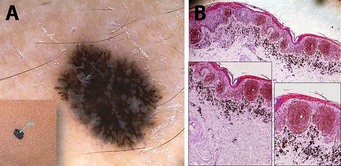
Figure 1.
Black-colored, flat lesion on the right thigh of an 11-year-old female child. (A) A superficial black network emerges under the dermatoscopic examination, overlying a diffuse bluish pigmentation. (B) Histopathology shows a junctional melanocytic lesion with focal areas of pigmented parakeratosis (black arrow) which explains the superficial black network seen in (A). The dense band of superficial dermal melanophages is thought to be responsible for the bluish background (HE, ×20). Pigmented spindle-shaped melanocytes predominate in well-demarcated junctional nests (insets, ×100 and ×200). [Copyright: ©2016 Pedrosa et al.]

