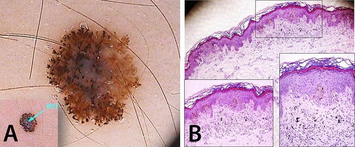
Figure 2.
Brown-colored lesion on the left arm of a 33-year-old female. (A) This atypical/multicomponent patterned lesion is dermatoscopically asymmetric typified by pseudopods irregularly distributed at the periphery and an atypical network attenuated at the right side. (B) Histopathology unveils a junctional asymmetric lesion with epidermal hyperplasia, hyperkeratosis and hypergranulosis exhibiting a focal infiltration of dermal melanophages responsible for the blue-whitish veil seen under dermoscopy (HE, ×40). Confluent epithelioid and spindle-shaped melanocyte nests are observed in insets (HE, ×100 and ×200). [Copyright: ©2016 Pedrosa et al.]

