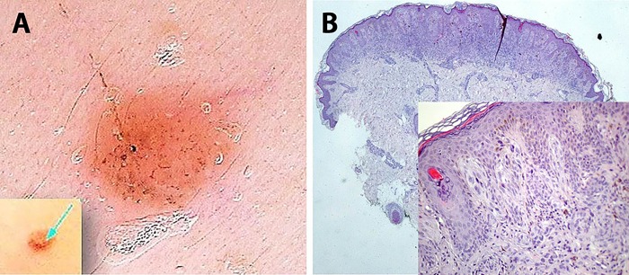
Figure 3.
A pink pale papule on the face of a 5-year-old female child. (A) A homogeneous pink-red pattern exhibiting dermatoscopically dotted and linear vessels and sparse pigmented globules not apparent at naked eye examination. (B) Histopathology shows a compound, symmetrical lesion with shallow depth, well-demarcated borders and a marked dermal inflammatory cell infiltrate in lower magnification (HE, ×40). Epithelioid and spindle-cell nests of melanocytes and sparse melanophages are observed, as well as scant pigment deposition (inset, ×200). [Copyright: ©2016 Pedrosa et al.]

