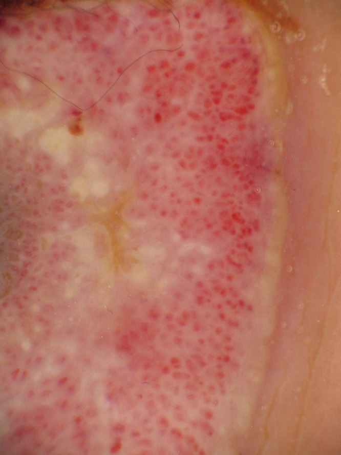
Figure 2.

Dermoscopy of condyloma lata. Red to milky-red globules, glomerular vessels and a whitish-pink network on the raised border. There is a yellowish structureless area at the periphery and multiple white, small, round structures in the center. [Copyright: ©2016 Ikeda et al.]
