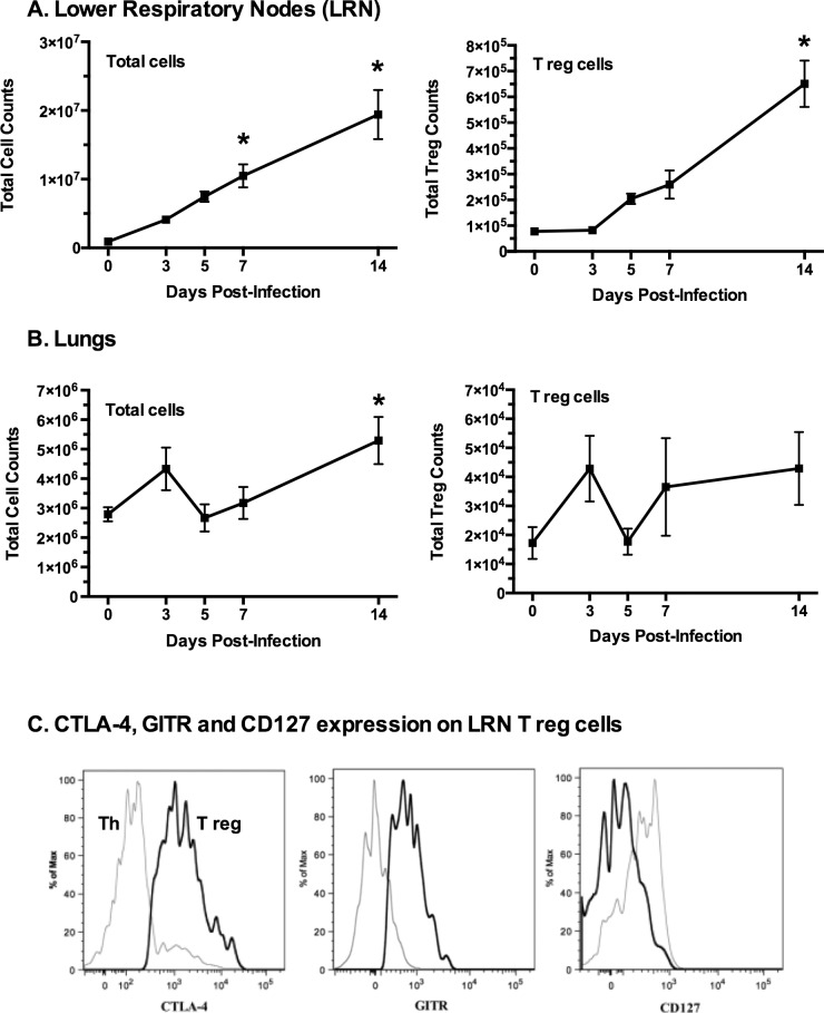
Fig 1. Treg (CD4+CD25+FoxP3+) cells increase after mycoplasma infection.
Mice were infected with M. pulmonis and sacrificed on days 0, 3, 5, 7, and 14 post-infection. Lymphocytes were isolated from (A) lower respiratory nodes (LRN) and (B) lungs. Treg cells were identified as CD4+CD25+FoxP3+ cells. An asterisk (*) indicates a significant difference (p ≤ 0.05) day 14 cell numbers versus day 0 cell numbers. Error bars represent the mean +/- SE (n = 9). (C) The majority of Treg cells in the LRNs were found to express high levels of CTLA-4 and GITR as compared to CD4+ effector T cells. In addition, Treg cells displayed little to no CD127 as compared to CD4+ cells. The black lines represent Tregs (CD4+CD25+FoxP3+) while gray lines represent CD4+CD25-FoxP3- Th cells. The histograms are from a representative LRN sample. Experiment was performed three times (n = 9).

