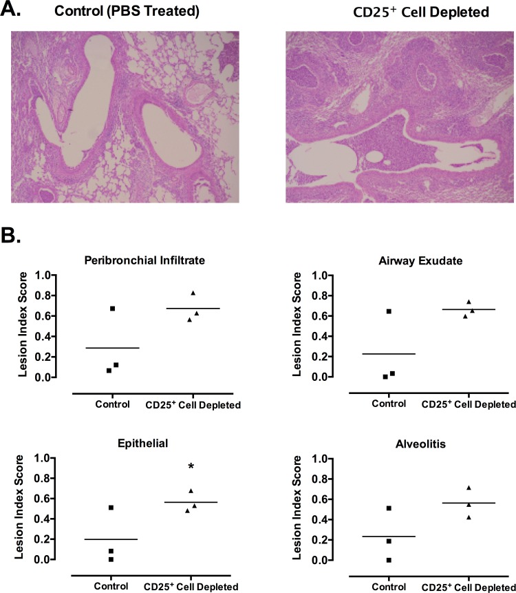
Fig 3. CD25+ cell-depleted mice develop more severe histopathology due to mycoplasma infection.
Mice were given anti-CD25 depleting antibody (CD25+ Cell Depleted) or PBS (Control) one day prior to infection with M. pulmonis, followed by antibody or PBS at day 6 post-infection. At 14 days post-infection, lungs were fixed for histology. (A) Representative sections from lungs of control (PBS treated) and Treg depleted infected mice are shown. (B) Lesion index scores for each of the characteristic lesions were determined, e.g. peribronchial and perivascular lymphoid hyperplasia or infiltration (peribronchial infiltrate); neutrophilic exudate in airway lumina (airway exudate); hyperplasia of airway mucosal epithelium (epithelial); and mixed neutrophilic and histiocytic exudate in alveoli (alveolitis). Individual data points and horizontal lines representing the means are shown (n = 3). An asterisk (*) represents a significant (P ≤ 0.05) difference. Overall, there was a significant (P ≤ 0.05) increase in the overall severity of lung lesions of mycoplasma disease when CD25+ T cells were depleted [PBS 0.24 +/-0.06; CD25+ Cell Depleted 0.62 +/- 0.06 (Mean +/- SE)].

