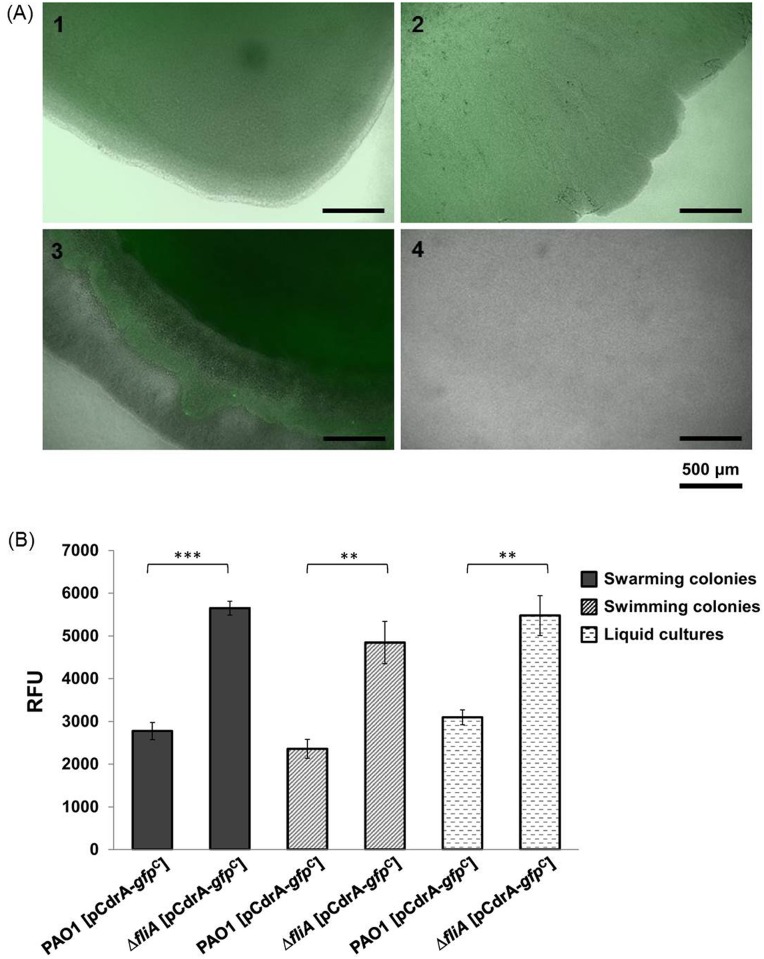
Fig 2. Intracellular concentration of c-di-GMP in P. aeruginosa wild-type PAO1 and fliA mutant.
(A) Fluorescence intensity of cells on the periphery of swarming (Panels 1 and 2) and swimming (Panels 3 and 4) areas are presented. Panels 1 and 3, PAO1 [pCdrA–gfpC]; 2 and 4, ΔfliA [pCdrA–gfpC]. (B) Relative fluorescence units of PAO1 [pCdrA–gfpC] and ΔfliA [pCdrA–gfpC] under different growth conditions.

