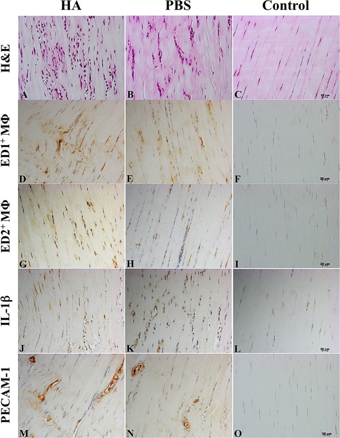
Fig 2. Immunohistochemistry images of histopathological changes after intratendinous injections.
Rat Achilles tendons stained with hematoxylin and eosin (H&E) (A-C), ED1+ macrophages (D-F), ED2+macrophages (G-I), IL-1β (J-L), and platelet endothelial cell adhesion molecule (PECAM-1) (M-O) on day 7 after an intratendinous injection ofhyaluronate (HA), phosphate buffered saline (PBS), or neither (Control: needle punctures only), respectively (from left to right). Magnification: 200x; bar = 20μm.

