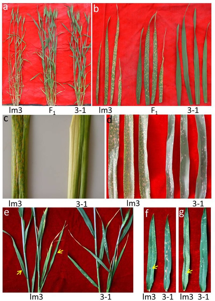
Fig 1. Phenotype of the lm3 mutant.
a Phenotype of the lm3 mutant, the wildtype (3–1) plants and their F1 progeny at two weeks after anthesis in the field. b Comparison of lesion symptoms in different leaves in the lm3 mutant, the wildtype (3–1) plants and their F1 progeny. For each line, the top fourth, top third, top second and flag leaves are presented from left to right. c Lesion symptoms on the leaf sheath of the lm3 mutant, and wildtype (3–1). d-g Enhanced resistance to powdery mildew. d Reaction of the lm3 mutant and wildtype (3–1) plant to Blumeria graminis f. sp. tritici (Bgt) under natural infection in the adult plant stage under field conditions in 2009. The flag leaves of lm3 plants were covered with yellow necrotic spots, but no spores of Bgt were visible, while many white and brown powdery mildew spores were present on the wildtype (3–1) plants. e Powdery mildew reaction under natural infection at the adult plant stage in the greenhouse in 2015. Only two instances of Bgt sporulation were observed on the older leaves of lm3 plants (yellow arrows), but many white powdery mildew spores were visible on the wildtype (3–1) plants. f and g Responses of the lm3 mutant and wildtype (3–1) plants to Bgt E18 in the growth chamber at the adult stage in 2015. Many white and brown spores of Bgt were observed on the flag leaves (f, right) and top second leaves (g, right) leaves of wildtype (3–1) plants, whereas only one spore (yellow arrows) was present on each of the corresponding leaves of the lm3 mutant (f and g, left), where necrotic spots were widely distributed. All the visible brown spots on lm3 leaves and sheath are necrotic lesions, except a few spores of Bgt indicated by yellow arrows on lm3 leaves (e, f and g), while most visible whitish spots on the leaves of wildtype (3–1) plants are spores of Bgt on the panels (d, e, f and g).

