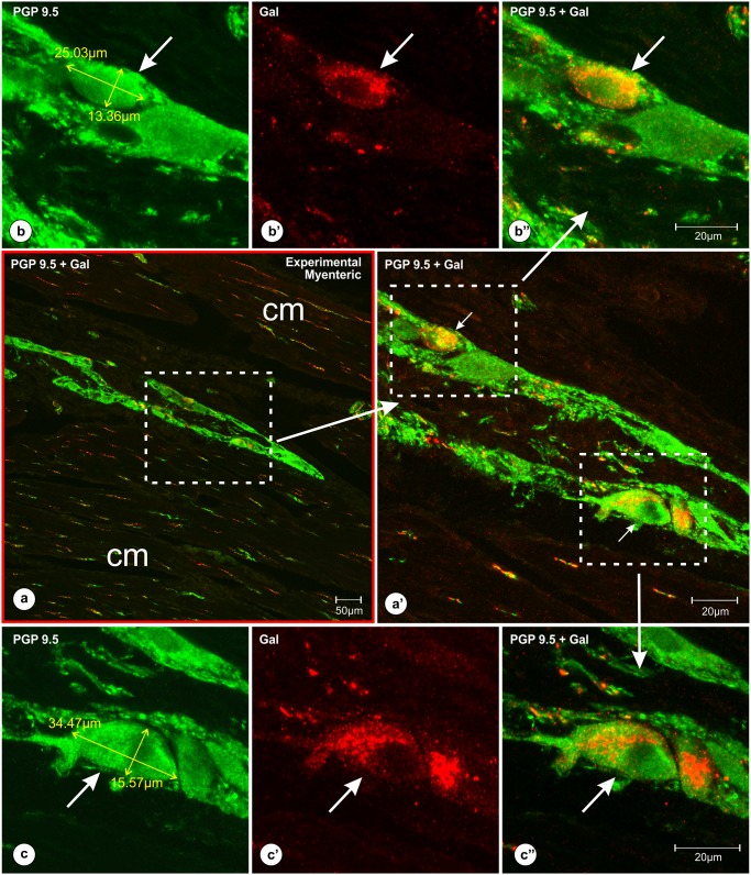
Fig 2. Localization of the myenteric plexus ganglia (containing Gal-immunoreactive perikarya) within the deep layers of the pyloric circular muscles.
Set of microphotographs at different magnifications of the pyloric wall cross-section comprising myenteric plexus ganglion with PGP 9.5/Gal-immunoreactive neurons. The section was taken from the experimental pig of the subgroup H and double-immunolabeled with antibodies against PGP 9.5 (b, c) and galanin (b’, c’) [pictures (a, a’, b”, c”) present overlap of both fluorescence channels]. Low magnification picture (a, red frame) shows a myenteric plexus ganglion localized deep within the pyloric circular muscle layers [cm]. Medium magnification picture (a’) of the selected area [dotted line boarder from the picture (a)] presents Gal-immunofluorescent perikarya (arrows). High magnification pictures (b, b’, b’, c, c’, c”) show medium (b, b’, b”) and large (c, c’, c”) in a diameter PGP9.5/Gal-immunoreactive perikarya (arrows). The characteristic pattern of Gal-immunoreactivity observed in the neurons (b’, c’) blurred the outlines of the perikarya, precluding accurate measurements of the cell bodies using red channel [Gal immunostaining]. Thus, all the measurements of Gal-immunoreactive nerve cells were performed using the green channel [PGP 9.5 staining (b, c)]. Scale bars are included in the pictures.

