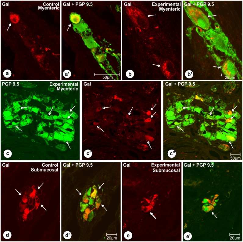
Fig 3. Typical characteristics (shapes, immunofluorescence) of Gal-immunoreactive perikarya.
Set of photomicrographs showing shapes and patterns of immunofluorescence observed in the typical Gal-positive (single arrows) myenteric (a, a’, b’ b’, c, c’, c”) and submucosal (d, d’, e, e’) perikarya of the pyloric orifice wall in the control (a, a’, d, d’) and experimental (b, b’, c, c’, c”, e, e’) pigs of the subgroup H. In both groups of the animals most of the myenteric neurocytes immunoreactive to galanin (arrows) were round (a, c’) or oval (b, c’) and expressed medium to strong immunoreactivity. In the experimental animals (b, b’, c, c’, c”) some of Gal-positive perikarya (double arrows) seemed to express weak immunofluorescence and/or were larger in a diameter. Most of the submucosal neurocytes immunoreactive to Gal (arrows) in the control (d, d’) and experimental (e, e’) animals were oval and measured about 19.45 ± 0.65 x 11.33 ± 0.38 μm. Scale bars are included in the pictures.

