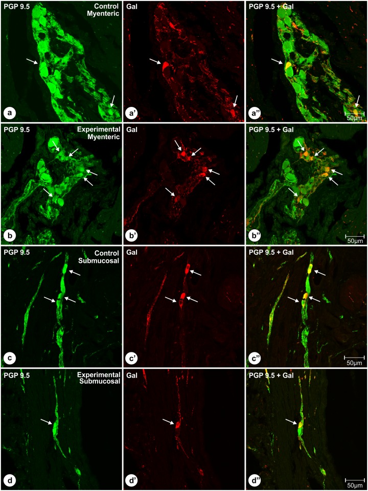
Fig 4. Double immunolabeled (PGP 9.5 and Gal) perikarya of the pyloric orifice wall.
Set of microphotographs showing sections of the pyloric orifice wall taken from the control and experimental pigs of the subgroup H and double-immunolabeled with antibodies against PGP 9.5 (a, b, c, d) and galanin (a’, b’, c’, d’). Some of the myenteric plexus perikarya (arrows) of the control (a, a’, a”) and experimental (b, b’, b”) animals simultaneously co-expressed immunoreactivity to PGP 9.5 (a, b) and galanin (a’, b’). The number of PGP 9.5+/Gal+ neurons was higher in the experimental animals and the difference was statistically significant. Some of the submucosal neurons (arrows) of the control (c, c’, c”) and experimental (d, d’, d”) animals simultaneously co-expressed immunoreactivity to PGP 9.5 (c, d) and galanin (c’, d’), and these percentages did not differ significantly between both groups of animals. Pictures (a”, b”, c”, d”) show the overlap of both fluorescence channels (PGP 9.5—green, Gal—red). Scale bars are included in the pictures.

