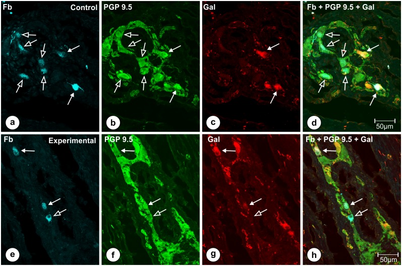
Fig 7. FB-positive neurons of the stomach antrum double-immunostained for PGP 9.5 and Gal.
Set of microphotographs showing stomach antrum sections with FB-positive neurons (a, e) from the animals of subgroup T and double-immunostained with antibodies against PGP 9.5 (b, f) and galanin (c, g). Some of FB-positive perikarya (solid arrows) in the control (a) and experimental (e) animals simultaneously co-expressed immunoreactivity to PGP 9.5 (b, f) and galanin (c, g), while the other traced neuronal somata (empty arrows) were devoid of galanin immunoreactivity. Differences in the number of FB+/PGP 9.5+/Gal+ neurons did not differ significantly between both groups of the animals. Pictures (d, h) show the overlap of all three fluorescence channels (FB—blue, PGP 9.5—green, Gal—red). Scale bars are included in the pictures.

