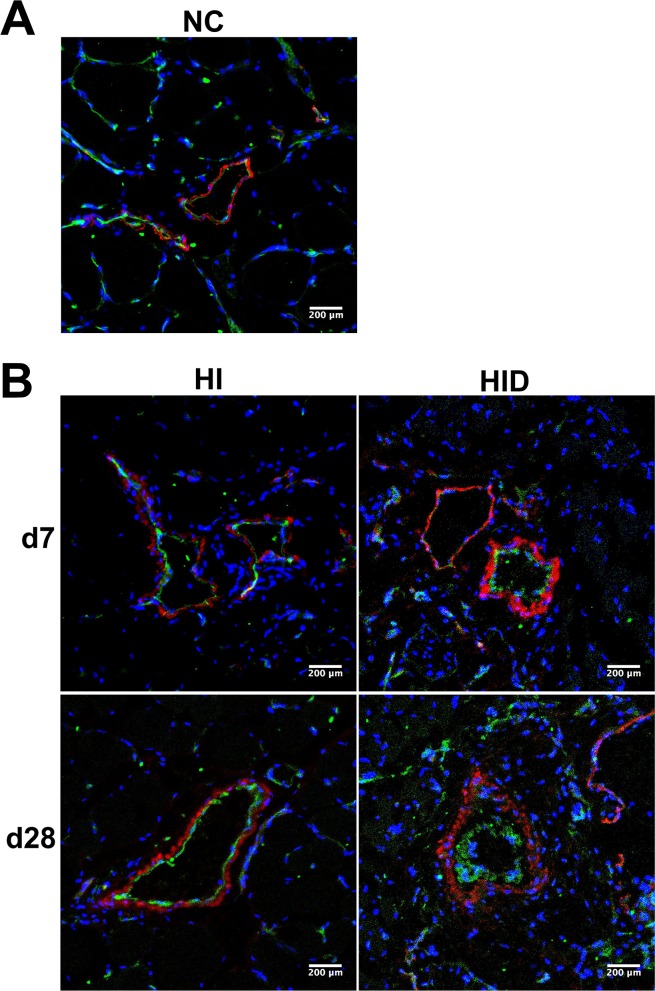
Fig 8. SMCs and ECs staining of collaterals in cross-sections of gracilis muscle.
Dual immunostaining of ECs with CD31 and SMCs with α-SM-actin in cross-sections of gracilis muscle in group NC, HI, HID. A thin layer of CD31 staining was observed in the collaterals of group NC (A) and HI on day 7 and 28 post-surgery, but the CD31 layer was much thicker on day 7 post-surgery and further thickened dramatically on day 28 post-surgery in group HID compared with group NC and HI (B). Similarly, the layer of α-SM-actin staining was thin in the collaterals of group NC (A). In group HI, the layer of α-SM-actin staining increased compared with group NC on day 7 and further increased slightly on day 28 post-surgery. In group HID, the staining layer increased significantly on day 7 and much further increased on day 28 post-surgery (B). CD31 indicates ECs (green), α-SM-actin indicates SMCs (red), and DAPI indicates nuclei (blue).

